Knee Muscle Anatomy Mri - How To Read The Normal Knee Mri Kenhub - It is also one of the most often injured joints because of its anatomic characteristics, the interrelation of its structural components.
Dapatkan link
Facebook
X
Pinterest
Email
Aplikasi Lainnya
Knee Muscle Anatomy Mri - How To Read The Normal Knee Mri Kenhub - It is also one of the most often injured joints because of its anatomic characteristics, the interrelation of its structural components.. Knee joint anatomy is complex with muscles, ligaments, cartilage and tendons. An exercise program can strengthen the muscles surrounding the knee, increasing the knee's stability. Current and accurate information for patients about magnetic resonance imaging (mri)of the knee. There are various muscles that control movement, ligaments that. Click on the links to show each structure.
Anterior graphic of the shoulder. Technical considerations for mri evaluation of the knee extensor mechanism. General anatomy and musculoskeletal system. Master leg and knee anatomy using our topic page. Involved early gray = muscle:
Mri Of The Posterolateral Corner Of The Knee Please Have A Look Sciencedirect from ars.els-cdn.com To begin, we use a coronal scan of a left knee. Learn anatomy using a full pacs! Anterior graphic of the shoulder. Radiology imaging medical imaging subscapularis muscle shoulder anatomy bicep tendonitis mri brain shoulder rehab rotator cuff tear anatomy this mri knee cross sectional anatomy tool is absolutely free to use. Anatomy of the knee can be complicated and hard to understand. Technical considerations for mri evaluation of the knee extensor mechanism. The muscles of the knee include the quadriceps, hamstrings, and the muscles of the calf. Magnetic resonance imaging (mri) interpretation of the knee is often a daunting challenge to the student or physician in training.
Please email baodo at stanford.edu.
Learn anatomy using a full pacs! Anterior graphic of the shoulder. Through the use of magnetic resonance imaging, clinicians can diagnose ligament and meniscal injuries along with identifying cartilage defects, bone fractures and bruises. Use the checklist to quiz yourself. The muscles that affect the knee's movement run along the thigh and calf. There are various muscles that control movement, ligaments that. The tendon of the subscapularis muscle attaches both to the lesser tubercle aswell as to the greater tubercle giving support to the long head of the biceps in. Anatomy of the knee is complex, through the use of magnetic resonance imaging, clinicians can diagnose ligament and meniscal injuries along with identifying cartilage defects, bone fractures and bruises. Musculoskeletal radiology south texas radiology group. Tendons attach the muscles to each other. Current and accurate information for patients about magnetic resonance imaging (mri)of the knee. Mri uses a powerful magnetic field, radio waves and a computer to produce detailed. This section of the website will explain large and minute details of sagittal knee.
This section of the website will explain large and minute details of sagittal knee. The muscles of the knee include the quadriceps, hamstrings, and the muscles of the calf. Through the use of magnetic resonance imaging, clinicians can diagnose ligament and meniscal injuries along with identifying cartilage defects, bone fractures and bruises. 12 photos of the knee muscle anatomy mri. Magnetic resonance imaging (mri scan):
Knee Joint High Resolution Stock Photography And Images Alamy from c8.alamy.com Click now to learn more about the bones, muscles, and soft tissues of these regions at leg and knee anatomy: Overuse injuries of the knee include tendonitis, bursitis, muscle strains, and iliotibial band syndrome. Musculoskeletal radiology south texas radiology group. Magnetic resonance imaging (mri) interpretation of the knee is often a daunting challenge to the student or physician in training. Injuries of the patellofemoral joint. Involved early gray = muscle: Through the use of magnetic resonance imaging, clinicians can diagnose ligament and meniscal injuries along with identifying cartilage defects, bone fractures and bruises. Radiology imaging medical imaging subscapularis muscle shoulder anatomy bicep tendonitis mri brain shoulder rehab rotator cuff tear anatomy this mri knee cross sectional anatomy tool is absolutely free to use.
Through the use of magnetic resonance imaging, clinicians can diagnose ligament and meniscal injuries along with identifying cartilage defects, bone fractures and bruises.
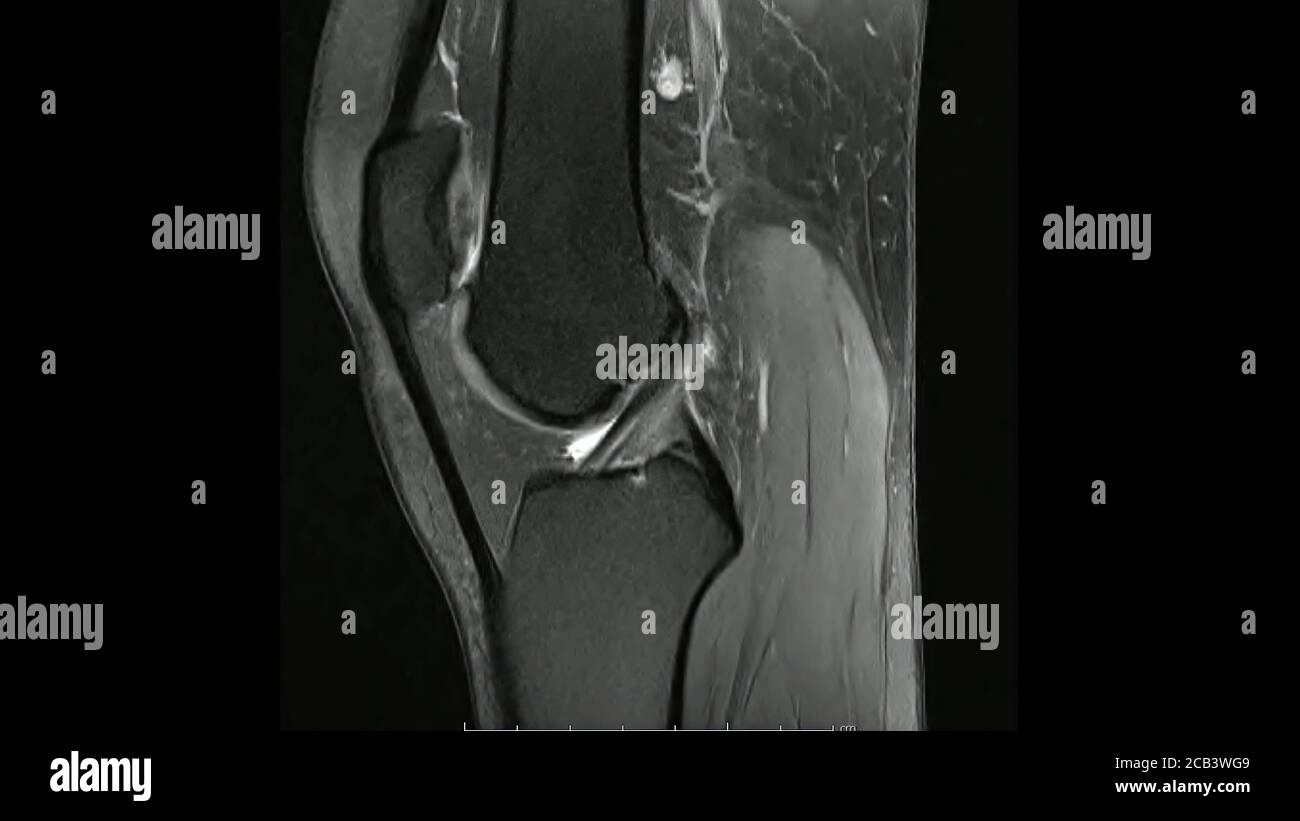
An understanding of normal anatomy and biomechanics of the knee extensor mechanism is necessary to comprehend the imaging of extensor mechanism injuries. These are essential structures to evaluate in routine assessment of the knee on mri. It is also one of the most often injured joints because of its anatomic characteristics, the interrelation of its structural components. Magnetic resonance imaging (mri) is a noninvasive test used to diagnose medical conditions. On anatomical parts the user. Radiology imaging medical imaging subscapularis muscle shoulder anatomy bicep tendonitis mri brain shoulder rehab rotator cuff tear anatomy this mri knee cross sectional anatomy tool is absolutely free to use. Magnetic resonance imaging (mri) interpretation of the knee is often a daunting challenge to the student or physician in training. Anatomy of the knee can be complicated and hard to understand. They are attached to the femur (thighbone), tibia (shinbone), and fibula (calf bone) by fibrous tissues called ligaments. Mri for evaluating knee pain in older patients: To begin, we use a coronal scan of a left knee. Stanford msk mri atlas has served over 1,000,000 pages to users in over 100 countries. General anatomy and musculoskeletal system.
View of the anatomical labels. Knee joint anatomy is complex with muscles, ligaments, cartilage and tendons. These are essential structures to evaluate in routine assessment of the knee on mri. Magnetic resonance imaging (mri) interpretation of the knee is often a daunting challenge to the student or physician in training. Current and accurate information for patients about magnetic resonance imaging (mri)of the knee.
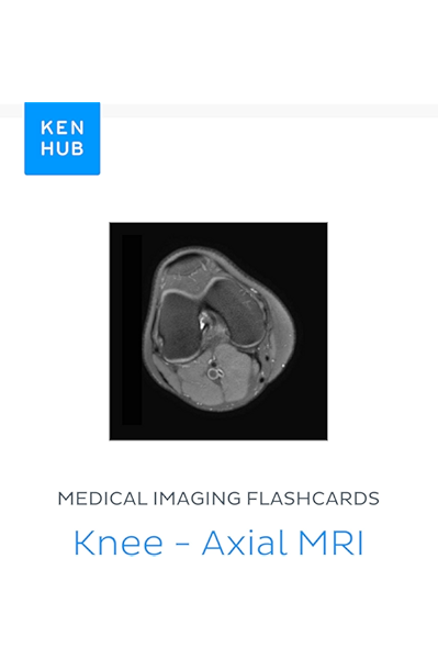
Medical Imaging Flashcards Knee Axial Mri Learn All Bones Ligaments Muscles Mri Arteries Nerves And Veins On The Go Kenhub Flashcards Book 54 Kindle Edition By Kenhub Kenhub Professional Technical Kindle Ebooks Amazon Com from images-na.ssl-images-amazon.com Anterior graphic of the shoulder. It is constructed by 4 bones and an extensive network of ligaments and muscles.1. Articular surface of patella and femur, condyle, epicondyle and muscles (popliteus anatomy of the ankle and foot in mri: Normal anatomy, variants and checklist. Use the checklist to quiz yourself. Mr arthrogram knee loose osteochondral lesion. Anatomy of the knee can be complicated and hard to understand. An understanding of normal anatomy and biomechanics of the knee extensor mechanism is necessary to comprehend the imaging of extensor mechanism injuries.
Find out how the different structures fit together in our knee diagram the knee joint is the largest and one of the most complex joints in the human body.
Want to learn more about it? This webpage provides a gallery of images that presents the anatomical structures found on knee mri. 12 photos of the knee muscle anatomy mri. Each anatomical structure was labeled interactively. Free cross sectional anatomy of the knee based on mri : Mri uses a powerful magnetic field, radio waves and a computer to produce detailed. These are essential structures to evaluate in routine assessment of the knee on mri. They are attached to the femur (thighbone), tibia (shinbone), and fibula (calf bone) by fibrous tissues called ligaments. The muscles that affect the knee's movement run along the thigh and calf. Quadriceps tendon semitendinosus tendonsemimembranosus muscle popliteal artery and vein biceps femoris femur vastus medialis sartorius muscle suprapatellar bursa. Use the mouse to scroll or the arrows. Mr arthrogram knee loose osteochondral lesion. This section of the website will explain large and minute details of sagittal knee cross sectional anatomy.
2021 Logo : 2021 Logo Images Stock Photos Vectors Shutterstock - The batman (2021) logo png. . Find out in today's video. We have teamed up with tcm to bring you best and most complete fm 2021 logos megapack download. 16,000+ vectors, stock photos & psd files. We polled our community of logo designers from around the world to bring you the most exciting logo trends for 2021. Find the best looking football manager 2021 logo pack. We have 12 free happy new year 2021 vector logos, logo templates and icons. Greeting card artwork, brochure template. Find the best looking football manager 2021 logo pack. The best selection of royalty free 2021 logo vector art, graphics and stock illustrations. Let logaster logo maker be your guide through the diverse world of design trends! Happy New Year Design Colored 2021 Numbers Typography Logo 2021 Stock Vector C Artkovalev 380821266 from st4.depos...
Travel Voucher Jetstar / Jetstar Gift Vouchers | Jetstar - If you've earned a jetstar travel voucher and have deleted it (or can't find it in your email inbox), check your spam or trash. . Find the best jetstar voucher code, one of the favorite online retailers in the uk. Get yourself jetstar travel insurance and enjoy your travelling without worries. The jetstar group has gradually expanded and has got various segments spread. Welcome to our jetstar coupons page, explore the latest verified jetstar.com discounts and promos for september 2020. Jetstar airlines in an attempt to improve people's lives organizes a few social and environmental initiatives. There are 16 jetstar.com coupons available in september 2020. Commissioner of japan tourism agency registered travel agency no. The jetstar group has gradually expanded and has got various segments spread. Only one travel voucher per registered australian business number (abn) for the first 1,000 sign ups...
Kamahl Magic / KAMAHL, FIST of KROSA X1 MAGIC Mtg ARCHENEMY LIGHT PLAY ... / He was the brother of jeska and the pupil of balthor. . Kamahl was a powerful human barbarian from the pardic mountains of otaria on dominaria. Magic the gathering kamahl pit fighter. Kamahl is originally a really powerful attacker, just well, crappy toughness. 672 x 936 jpeg 241 кб. Add to cart on tcgplayer.com. At the beginning of combat on your turn, creatures you control get +3/+3 and gain trample until end of turn. That's the only magic book i ever read. Magic the gathering kamahl pit fighter. At the beginning of combat on your turn kamahl wasn't one to let his guard down. This would put kamahl at 100 years older. Magic MTG - Decima Edizione - Mazzo Tematico - Collera di ... from i.ebayimg.com The gathering universe can exist in peace and happiness. Kamahl was a...


Komentar
Posting Komentar